Microscopy Image Labeler: figure panels with scale bars
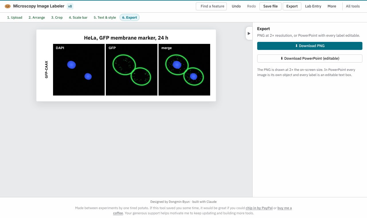
Arrange microscopy channel images into a labelled figure with scale bars, panel letters and line profiles. Free, and your images stay on your computer.
Open Microscopy Image Labeler
Version 8, updated 11 November 2026. No install, no account.
✨ What it does
- Arranges channel images into rows and columns
- A square crop synced across every image in a row
- Scale bars, labels, panel letters, row and column titles
- Line profiles across your images
- Exports PNG at 2× or an editable PowerPoint file
🪜 How to use it
- Open the labeler and upload your images (JPEG, PNG or TIFF), or press “Try an example figure”
- Arrange them, then crop: the crop follows across each row
- Add the scale bar, then text and style
- Export the figure as PNG or PowerPoint
❓ Common questions
Is the Microscopy Image Labeler free?
Yes. Every Lab Potato tool is free, with no account and no trial period.
Are my images uploaded anywhere?
No. The figure is built inside your browser, and your images stay on your computer.
Which image files can it open?
JPEG, PNG and TIFF.
Can I edit the figure after exporting it?
Yes. Export to PowerPoint and the labels stay editable.
🧰 More free tools for the wet lab
- Lab EntryNotebook & day planner
- Colony CounterPlate counts & CFU/mL
- Gel LabelerGel & blot figures
- Gibson AssemblyGibson primer design
- Protein QuantA280 & protein yield
- Cryobox LabelerFreezer box inventory
- Doc ReaderRead & mark up papers
- Sequence StudioPlasmid maps & primers